Tuberculoid Leprosy
Mohsen Esfandbod, M.D.
N Engl J Med 2011; 364:1657April 28, 2011
Mohsen Esfandbod, M.D.
Tehran University of Medical Sciences, Tehran, Iran
sfandbod@sina.tums.ac.ir
N Engl J Med 2011; 364:1657April 28, 2011
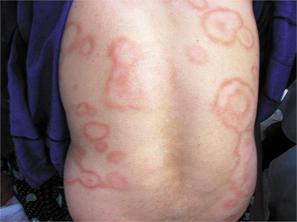
A 30-year-old woman presented with a 4-month history of a rash on her back. The physical examination revealed multiple large, annular, hypopigmented, atrophic macules with well-defined, erythematous, raised borders. The lesions were hairless, hypohidrotic, and anesthetic and had developed at a slow and progressive pace. There was no peripheral-nerve enlargement.
Histopathological analysis of a skin-biopsy specimen revealed well-developed epithelioid granulomas, lymphocytes, and Langerhans' cells surrounding neurovascular structures within the papillary dermis. Dermal nerves were swollen and destroyed. No acid-fast bacilli were detected on modified Ziehl–Neelsen staining. On the basis of clinical and histologic findings, the condition was diagnosed as tuberculoid leprosy. The clinical manifestations of leprosy depend on the nature of the host's immune response to infection with Mycobacterium leprae and range from lepromatous leprosy (uncontrolled replication with nerve damage from high-titer infection) to tuberculoid leprosy (nerve and organ damage predominantly from the host granulomatous immune response). The patient had no known contact with anyone with leprosy, which has a prevalence of less than 1 in 100,000 in Iran. She was treated with a 6-month course of rifampin and dapsone with nearly complete clearing of the skin lesions.
Histopathological analysis of a skin-biopsy specimen revealed well-developed epithelioid granulomas, lymphocytes, and Langerhans' cells surrounding neurovascular structures within the papillary dermis. Dermal nerves were swollen and destroyed. No acid-fast bacilli were detected on modified Ziehl–Neelsen staining. On the basis of clinical and histologic findings, the condition was diagnosed as tuberculoid leprosy. The clinical manifestations of leprosy depend on the nature of the host's immune response to infection with Mycobacterium leprae and range from lepromatous leprosy (uncontrolled replication with nerve damage from high-titer infection) to tuberculoid leprosy (nerve and organ damage predominantly from the host granulomatous immune response). The patient had no known contact with anyone with leprosy, which has a prevalence of less than 1 in 100,000 in Iran. She was treated with a 6-month course of rifampin and dapsone with nearly complete clearing of the skin lesions.
Tehran University of Medical Sciences, Tehran, Iran
sfandbod@sina.tums.ac.ir


Komentar
Posting Komentar
mampir comment dulu sodara..